PRODUCTImage scanner for electrophoretic gels
GELSCAN
GELSAN

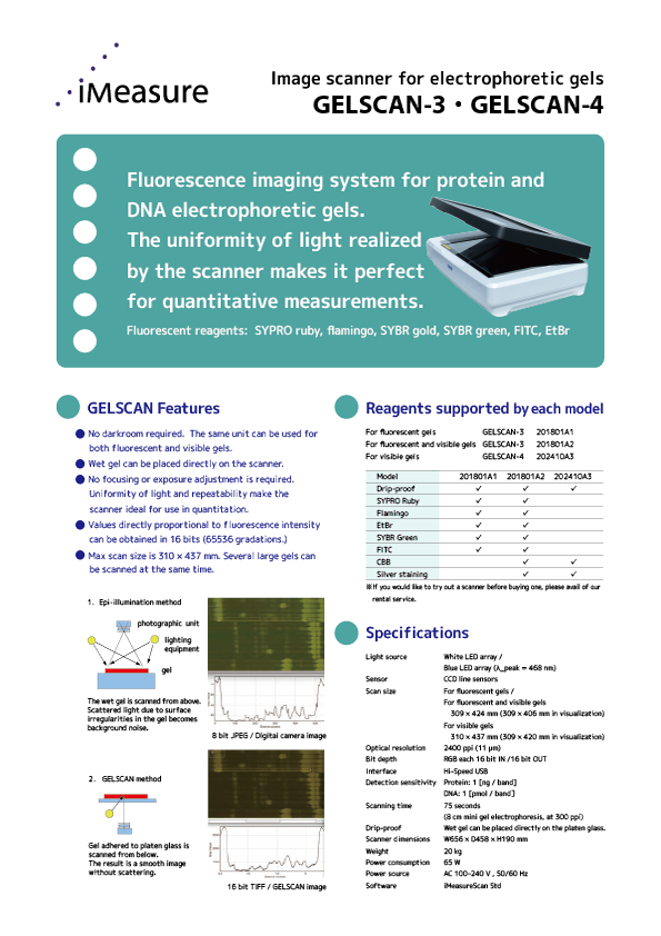
Fluorescence imaging system for protein and DNA electrophoretic gels.
The uniformity of light realized by the scanner makes it perfect for quantitative measurements.
Application examples
| Protein analysis.
| Improvement of plant varieties.
| Confirmation of DNA. ...etc.
GELSCAN SERIES LIST
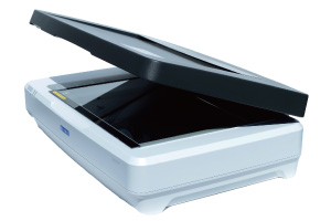
GELSCAN-4[For visible gels]
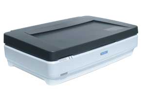
GELSCAN-3[For fluorescent gels,For fluorescent and visible gels]
![GELSCAN-2[End of sale]](/img/gelscan2.jpg)
GELSCAN-2[End of sale]
[Sales period]
November 22, 2014 - March 13, 2018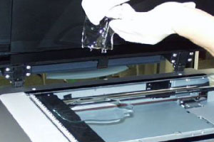
GELSCAN[End of sale]
[Sales period]
December 10, 2007 - November 21, 2014
SPECIFICATIONS
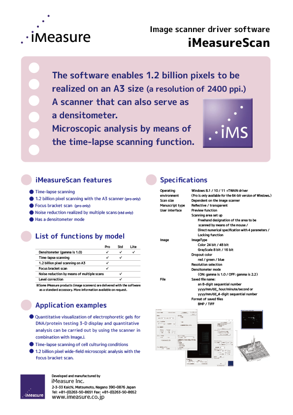
| Light source | White LED array / Blue LED array (λ_peak = 468 nm) |
|---|---|
| Sensor | CCD line sensors |
| Scan size | For fluorescent gels,For fluorescent and visible gels: 309 × 424 mm (309 × 406 mm in visualization) For visible gels: 310 × 437 mm (309 × 420 mm in visualization) |
| Optical resolution | 2400 ppi (11 μm) |
| Bit depth | RGB each 16 bit IN /16 bit OUT |
| Interface | Hi-Speed USB |
| Detection sensitivity | Protein: 1 [ng / band] DNA: 1 [pmol / band] |
| Scanning time | 75 seconds(8 cm mini gel electrophoresis, at 300 ppi) |
| Drip-proof | Wet gel can be placed directly on the platen glass. |
| Scanner dimensions | W656 × D458 × H190 mm |
| Weight | 20 kg |
| Power consumption | 65 W |
| Power source | AC100-240V,50/60Hz |
| Software | iMeasureScan Std |
| OS |
GELSCAN-3: TWAIN GELSCAN-4: TWAIN |
MODELS
| Product name | Scanner models | Function | Supported manuscripts | Specification |
|---|---|---|---|---|
GELSCAN-3 |
201801A1 |
Electrophoresis gel stain image scanner |
Visualization: luminescent gels |
[Luminescent stain] |
GELSCAN-3 |
201801A2 |
Electrophoresis gel stain and visualization image scanner |
Visualization: luminescent gels |
[Luminescent stain] |
GELSCAN-4 |
202410A3 |
Electrophoresis Gel Visualization Image Scanner |
Visualization: Reflective manuscript |
Drip-proof model. |
*GELSCAN and iMeasureScan (Windows 10/11, 32bit/64bit compatible) are included as standard accessories.
Option / Customize
| Product name | Summary | |
|---|---|---|
| Customize | Excitation / emission wavelength customization. |
The excitation wavelength and fluorescence wavelength can be customized by changing the transmission filter on the light source side, the cut filter on the sensor side, and the emission center wavelength of the light source. (Estimate separately for price and delivery date.) |
| Option | Unit |
By attaching to a fluorescent model, it is possible to scan transmitted originals (CBB / silver stain). * Fluorescent / visible (201801A2) model is attached as standard. |
| Option | Carrying case |
Made of aluminum frame + FRP. |
FEATURES
01No darkroom required.
The same unit can be used for both fluorescent and visible gels.
02Drip-proof.
Wet gel can be placed directly on the scanner.
03The uniformity of light realized by the scanner makes it perfect for quantitative measurements.
No focusing or exposure adjustment is required. Uniformity of light and repeatability make the scanner ideal for use in quantitation.
0465536 gradations.
Values directly proportional to fluorescence intensity can be obtained in 16 bits (65536 gradations.)
05Max scan size is 310 × 437 mm.
Several large gels can be scanned at the same time.
APPLICATION EXAMPLES
-
DNA '3'-end modification' FITC
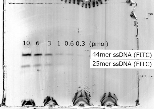
[Scan condition]
Electrophoresis Gel Stain Imager GELSCAN Optical resolution:300ppi (pixel per inch, 85μm) Gradation:16-bit gray scale
[Target]
44mer ssDNA(FITC). 25mer ssDNA(FITC) Density: 10, 6, 3, 1, 0.6, 0.3 [pmol] 3'-terminal FITC modification -
fluorescence stain:SYBR Green
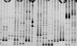
Images from the Illuminator + Digital Still Camera System
[Scan condition]
Scanning time:63 sec Optical resolution:300ppi (pixel per inch, 85μm) Gradation: 16-bit Green channel
[Source]
Nagano Vegetable and Ornamental Crops Experiment Station -
fluorescence stain:EtBr
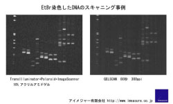
[illuminator + Polaroid Camera] VS GELSCAN
[Source]
Nagano Vegetable and Ornamental Crops Experiment Station -
fluorescence stain:SYBR Gold
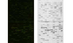
[Scan condition]
Gel size: 265 x 435mm(2 sheets) Scanning time:7 minutes Optical resolution:300ppi, Gradation: 48-bitColor (16 Million pixels) -
Protein fluorescence staining: Flamingo
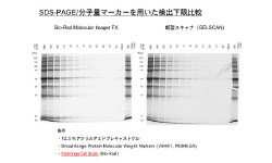
BIO-RAD Molecular Imager FX vs GELSCAN
-
Protein fluorescence staining: SYPRO Ruby
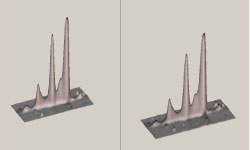
[Experimental material]
Creating SYPRO Ruby calibrations
DEVELOPER
Sales Agent
(1) NIHON EIDO.Corp
3F, Mitomi Bld., 3-13-3, Hongo, Bunkyo Ku, Tokyo To, 113-0033, Japan
+81-3-3818-1691 |
+81-3-38-18-6967 |
//www.nihon-eido.jp/newarrival/page09.html/a>
(2) Berthold Japan K.K.
3-6-24 Tenjinbashi, Kita-ku, Osaka-shi, Osaka 530-0041
//www.berthold-jp.com/products/lifescience/imagingscanner.html
Person in charge
iMeasure Inc. Yoshinori KANESAKA
[Collaborators]
TOWA ENVIRONMENT SCIENCE CO.,LTD. / IDEA Consultants, Inc. ken OOFUSA
 ACCESS
 CONTACT US